
This content is reviewed for medical accuracy, compliance with current guidelines and patient safety. expert supervision has been subjected.
Current scientific literature and national clinical guidelines and in-center practical knowledge.



Most people diagnosed with diabetes are advised on blood sugar monitoring, nutrition, and foot care. Eye exams, however, are often left at the bottom of the list and put off with the excuse, “There’s nothing wrong with my vision.”.
This delay overlooks the most important aspect of diabetes as it relates to the eyes. Most of the changes affecting the eyes show no symptoms in the early stages. By the time a person notices something, the condition may already be well advanced.
The following explains how diabetes affects different parts of the eye, how quickly certain conditions develop, why the screening schedule is structured this way, and what measures are truly effective in reducing the risk.
Straight Answer: Diabetes can affect the retina, the lens, and intraocular pressure. The most common and serious complication is diabetic retinopathy, which develops due to damage to the blood vessels of the retina. In the early stages, it causes no symptoms, and visual acuity may be measured as normal. For this reason, regular fundus examinations are necessary starting from the date of diagnosis, even if there are no vision-related complaints.
How does diabetes affect the eyes?
The effects of diabetes on the eye do not constitute a single disease. Prolonged high blood sugar levels affect different structures of the eye through different mechanisms, and these conditions can coexist in the same person.
The most significant effect involves the small blood vessels in the retina. Over time, high blood sugar weakens the walls of these blood vessels; the vessels begin to leak, some become blocked, and a portion of the retina is no longer adequately nourished. This condition is called diabetic retinopathy and is the most common eye-related complication of diabetes.
The second effect is on the eye's lens. Because the biochemical balance within the lens is directly affected by blood sugar levels, cataracts in people with diabetes can develop at an earlier age and progress more rapidly.
The third group consists of less well-known effects. These include increased intraocular pressure, symptoms of dryness due to disruption of the tear film, and slowed healing of the corneal surface. In rare cases, the nerves that control the eye muscles may be temporarily affected, leading to double vision.
The most critical information: There are no symptoms in the early stages
The most dangerous aspect of diabetic retinopathy is that it begins silently. When changes in the retina begin, the person usually feels nothing, and visual acuity may still be measured as normal.
For this reason, the assumption that “my vision is fine, so I don’t need an eye exam” does not apply to diabetes. The onset of vision problems may indicate that the disease has already progressed beyond its early stages. The absence of symptoms is not a guarantee of good health; it merely reflects a lack of information.
This is why the screening schedule is based on the date of diagnosis rather than on symptoms. In diabetes care, an eye exam is not a response to a complaint but a pre-scheduled checkup; the appointment is made not when symptoms arise, but when the scheduled date arrives.
What is diabetic retinopathy, and how does it progress?
Diabetic retinopathy is a condition that develops as a result of damage to the small blood vessels that supply the retina over the course of many years. The retina is the layer of nerve tissue at the back of the eye where images are formed; when the blood supply to this layer is impaired, vision can be permanently affected.
The process is gradual and is generally examined in two main stages. In the early stage, changes in the blood vessels are confined to the retina. In the advanced stage, however, the eye begins to form new blood vessels to compensate for the areas that are not receiving adequate blood supply, and the main problems arise at this stage.
There is a third condition that can develop independently of these two stages. Fluid buildup in the central area of the retina can impair central vision, regardless of the stage of the disease.
Early stage: onset of vascular changes
During this period, small bubbles form in the blood vessel walls, and pinpoint hemorrhages and deposits caused by leakage may be observed. These changes remain confined to the retina, and vision is not affected in most people.
The entire early stage can be detected only through a fundus examination. Since there are no symptoms that a person can notice, diagnosis at this stage depends entirely on regular screening. This is the stage when the disease is easiest to manage and most often goes undetected.
Advanced stage: new blood vessel formation
When the number of areas in the retinal layer that are not receiving adequate blood supply increases, the eye attempts to compensate by generating new blood vessels. This is a repair mechanism, but the outcome is not favorable. The newly formed blood vessels are structurally weak, run irregularly, and bleed easily.
If bleeding occurs in the eye’s transparent inner cavity, vision may suddenly become impaired. The person may describe seeing dark spots, dense floating objects, or a veil in front of their eyes; this condition requires immediate evaluation.
Over time, the connective tissue that forms around these blood vessels can pull on the retinal layer, causing it to detach. In addition, new blood vessel formation can affect the front of the eye, leading to an increase in intraocular pressure. Although treatment options are available in advanced stages, it is not always possible to fully restore lost vision.
Diabetic macular edema
The macula is the area located at the center of the retina that enables sharp vision for tasks such as reading and recognizing faces. The accumulation of fluid in this area due to leaking blood vessels is called diabetic macular edema.
This condition can also occur in the early stages of the disease and directly affects central vision. The person may describe seeing the centers of letters disappear, straight lines appearing wavy, or having difficulty reading. Macular edema is the most common cause of vision loss, regardless of the stage of retinopathy.
Situations That Require Seeking Medical Attention Without Delay
The following symptoms are not expected in a person with diabetes and should be evaluated without delay:
- If your vision has noticeably decreased within a few hours or a few days,
- If you suddenly notice an increase in floating objects, dark spots, or soot-like images in your field of vision,
- If a patient describes a curtain-like obstruction or a dark spot in part of their visual field
- If you experience sudden flashes of light
- If the centers of the letters disappear while you're reading, or if straight lines appear wavy,
- If you have eye pain, noticeable redness, or a headache accompanied by nausea
- If you start seeing double
None of these findings should be ignored with the thought, “My blood sugar is high, but it will go back to normal.” In cases involving the retina, the time lost may be irreparable.
Why do cataracts develop earlier in people with diabetes?
A cataract is the loss of transparency in the eye's natural lens and can occur in anyone as they age. In people with diabetes, this process is expected to begin earlier and progress more rapidly.
The reason has to do with the lens’s own structure. The lens is a tissue that reacts directly to changes in blood sugar; prolonged high blood sugar levels disrupt the balance within the lens and accelerate the loss of transparency. For this reason, cataracts can be detected in people with diabetes as early as their 40s and 50s.
Cataracts also have an indirect effect. A clouded lens makes it difficult to evaluate the back of the eye, and in such cases, monitoring the retina may be compromised; Symptoms of Cataracts and the Diagnostic Process For this reason, diabetes management is tracked under a separate heading.
The Relationship Between Diabetes and Glaucoma
People with diabetes are more likely to experience elevated intraocular pressure than the general population. For this reason, intraocular pressure is routinely measured during eye exams.
In advanced retinopathy, however, a different mechanism comes into play. New blood vessels that form in the front of the eye can block fluid circulation, raising pressure, and this condition can lead to a situation that is difficult to treat. Symptoms and Monitoring of Intraocular Pressure This is another topic that is monitored in people with diabetes.
Why do blood sugar fluctuations cause blurred vision?
A condition that people with diabetes often experience but is little known is temporary blurred vision when blood sugar levels fluctuate. This condition is often mistaken for a cataract.
This mechanism is related to the lens’s water content. When blood sugar rises, the fluid balance within the lens changes, the lens’s refractive power shifts, and vision can become noticeably impaired within a few days. Once blood sugar levels stabilize, this change reverses.
For this reason, having your prescription checked during a period when your blood sugar has just been stabilized will not yield accurate results. To ensure the prescription stabilizes, it is necessary to wait for blood sugar levels to remain stable for a while. Otherwise, the glasses you receive will no longer be suitable after a short time.
Common Mistakes
“The belief that ”as long as my blood sugar is under control, I won’t have any eye problems” is common but misleading. Good glycemic control significantly reduces the risk, but it does not eliminate it. Furthermore, the duration of diabetes is at least as important a factor in the development of retinopathy as blood sugar levels; even if blood sugar is well-controlled in a person who has had diabetes for a long time, the screening interval is not shortened.
Two less-discussed symptoms of diabetes: dryness and delayed healing
The effects of diabetes on the eye are not limited to the retina and the lens. Dryness caused by a disrupted tear film is more common in people with diabetes than in the general population and manifests as a stinging, burning sensation, a feeling of having sand in the eyes, and fluctuating blurred vision throughout the day. This symptom is often attributed to fatigue and goes unreported.
The second point is that healing of the corneal surface may be delayed. A minor scratch or superficial irritation in the eye may take longer than expected to heal in people with diabetes, and the risk of infection may increase; therefore, persistent redness and discomfort in the eye should be evaluated without delay.
A common feature of these two complaints is that they are often taken lightly. Neither involves the retina, nor do they pose a risk of vision loss. However, they significantly affect daily comfort and can be easily assessed during an eye exam; therefore, it is appropriate to bring them up rather than dismissing them as “insignificant.”.
When and how often should an eye exam be performed?
Eye screenings for diabetes are scheduled based on the date of diagnosis, not in response to specific symptoms. This is the key difference that sets diabetes care apart from many other medical conditions.
The purpose of the screening is not to measure vision. The goal is to detect changes in the retina that have not yet caused symptoms and, if necessary, refer the patient for further evaluation. Measuring visual acuity is only one part of this screening and is not sufficient on its own.
The screening interval varies depending on the findings. If no findings are identified during the examination, the interval can be extended; if early-stage findings are detected, follow-up visits are scheduled more frequently and the follow-up plan is revised.
The interval may also be shortened during periods when blood sugar control is impaired, blood pressure rises, or changes in kidney function are observed. For this reason, the eye follow-up schedule is not a fixed calendar. Internal Medicine It is updated based on this assessment and reconfigured according to the individual's overall profile.
In Type 2 diabetes
In type 2 diabetes, an eye exam is scheduled as soon as the diagnosis is made. This is because type 2 diabetes often goes unnoticed for years, and by the time it is diagnosed, changes in the retina may have already begun.
The frequency of follow-up visits is determined based on the results of the initial evaluation. Type 2 diabetes In follow-up care, eye exams are just as routine as blood tests. Skipping an eye exam while regularly monitoring blood sugar levels indicates that follow-up care is not being properly conducted.
In Type 1 diabetes
In Type 1 diabetes, the time of diagnosis is usually close to the onset of the disease. For this reason, the first eye exam is scheduled for a certain period after diagnosis; the timing and subsequent intervals are determined by the physician.
For individuals diagnosed during childhood and adolescence, follow-up care continues throughout the growth period. Hormonal changes during adolescence can make it difficult to control blood sugar levels. This is the period when follow-up care is most often neglected, yet it is when it is needed the most.
Pregnancy and Pregnancy Planning
Pregnancy is a unique period that can accelerate the progression of diabetic retinopathy. For this reason, women with diabetes who are planning to become pregnant should have an eye exam before becoming pregnant.
Follow-up continues throughout the pregnancy, with the frequency determined by the doctor; evaluations also continue during the postpartum period.
This tracking, Gynecology and Obstetrics It is managed in conjunction with monitoring. Gestational diabetes, which develops during pregnancy, is a different condition and is evaluated separately in terms of eye monitoring.
What happens during a fundus exam?
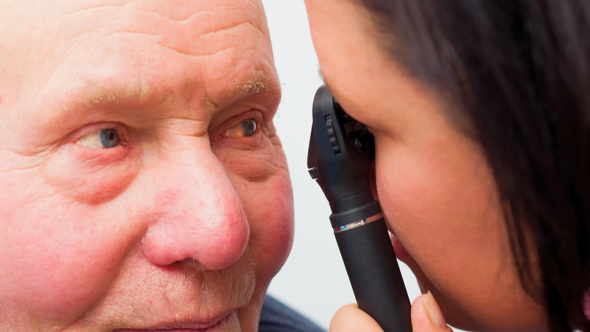
To examine the retina, the pupil must be dilated. Eye drops are used for this purpose, and it usually takes about half an hour for them to take effect.
While the eye drops are taking effect, it becomes difficult to read up close, and sensitivity to light increases. This effect lasts for a few hours. For this reason, it is recommended that you come to your appointment without driving and bring sunglasses with you.
During the examination, the retina, blood vessels, optic nerve head, and macula are evaluated separately. Visual acuity is measured and intraocular pressure is checked during the same session; if deemed necessary, the patient is referred for further imaging.
How is treatment planned once a finding is identified?
The detection of findings during a fundus examination does not necessarily mean that eye treatment will begin immediately. The approach is determined based on the stage of the disease and the condition of the macula.
In the early stages, most people do not require any eye-related procedures. At this stage, the primary treatment involves controlling blood sugar, blood pressure, and blood lipids; as for the eyes, the focus is on shortening the follow-up intervals and monitoring any changes. The benefits of metabolic control at this stage are significantly greater than those in later stages.
In advanced stages and in cases of macular edema, treatment options that target the eye directly come into play. These options include intravitreal injections, laser treatments, and, in some cases, surgery; the appropriate treatment is determined based on the type and extent of the findings. Since these procedures require advanced evaluation and specialized equipment, the patient is referred to an appropriate center once the condition is diagnosed. Timely referral is one of the factors that influence the outcome.
Expectation management becomes particularly important at this stage. The primary goal of treatments administered in the advanced stages is not to restore lost vision, but to halt progression and preserve existing vision; while some patients may experience an improvement in vision, this is not the goal itself. This is where it becomes most evident why early detection is so critical.
What can be done to reduce the risk?
Factors that reduce the risk of developing diabetic retinopathy are well known, and most of them are already part of routine diabetes care.
Long-term blood sugar control is at the top of this list. Keeping the long-term average blood sugar level within the target range reduces both the likelihood of developing retinopathy and the rate at which it progresses. The target value varies from person to person and is determined by the doctor.
Blood pressure control ranks second, and its importance is often underestimated. Since high blood pressure increases the strain on the retinal blood vessels, controlling blood pressure can be just as critical for eye health as controlling blood sugar; managing blood lipids and quitting smoking are also on the same list.
Nutrition and physical activity are an integral part of this plan. A personalized plan must be developed jointly by a dietitian and a doctor; general programs found online are not a safe starting point for managing diabetes. Nutrition and Dietetics This assessment is crucial for achieving sustainable blood sugar control.
Finally, monitoring itself is a risk-reduction method. Regular eye exams do not prevent the disease from developing, but they do allow for its detection at an early stage; when the condition is detected early, treatment options are broader and outcomes are better.
Questions to Ask Your Doctor
Asking these questions during your eye exam will make follow-up easier:
- Are there any findings in my endothelium, and if so, at what stage?
- When should I have my next eye exam?
- Do I have edema in my macula?
- If there’s a change in my blood sugar or blood pressure levels, should I schedule my eye exam sooner?
Why are eye, foot, and kidney checkups conducted together in diabetes?
The long-term effects of diabetes share a common underlying cause: damage to small blood vessels. The blood vessels in the retina, as well as those in the kidneys and feet, are affected by the same process.
The practical implication of this is as follows: findings in one organ serve as a warning sign for the others. In a patient with noticeable findings in the reticular layer, it is standard practice to review kidney function and examine the feet. The reverse is also true.
In the context of the foot, this monitoring involves assessing circulation and sensation, as well as tracking the risk of wounds. In individuals with existing wounds, this process becomes a separate topic and chronic wound Management steps in.
In practice, this means that diabetes management cannot be confined to a single appointment. Eye exams, foot exams, and blood tests are scheduled at different intervals, and the results of one may affect the frequency of the others. Diabetes management is therefore not the responsibility of a single specialty but rather the coordinated conduct of several interconnected evaluations.
Frequently Asked Questions
I have no problems with my vision—do I still need an eye exam?
Yes. The early effects of diabetes on the retina do not cause symptoms, and visual acuity may be measured as normal. The purpose of the examination is not to evaluate symptoms, but to detect changes that have not yet caused symptoms.
Can diabetic retinopathy be reversed?
Some of the symptoms detected in the early stages may remain stable or even improve with proper management of blood sugar and blood pressure. However, the structural changes and vision loss that occur in the advanced stages cannot always be reversed. This is why early detection is so important.
My blood sugar has stabilized, but my vision has become blurry—is this normal?
Rapid changes in blood sugar levels can temporarily affect the refractive power of the lens, and this blurred vision usually resolves once blood sugar levels stabilize. An evaluation is necessary if the blurred vision persists, occurs in only one eye, or is accompanied by new symptoms.
Will I go blind because of diabetes?
Diabetes is one of the preventable causes of vision loss. With regular screening and timely referral, the majority of advanced cases can be prevented. The key factor here is how early the condition is detected. Having diabetes does not necessarily mean you will experience vision loss; however, failing to monitor the condition significantly increases the risk.
Do I need to use eye drops?
There is no eye drop treatment for diabetic retinopathy. Eye drops may be recommended for accompanying conditions such as dry eye, but they do not affect the condition affecting the retina.
I was recently diagnosed with diabetes. Should I wait to have an eye exam?
In Type 2 diabetes, the first eye exam is scheduled at the time of diagnosis, as the disease may have gone unnoticed for a long time prior to diagnosis. There is no reason to wait; the first exam also serves as the baseline against which subsequent follow-ups will be compared.
What should I bring with me when I go to my appointment?
A list of the medications you are taking, your current blood sugar levels and long-term average blood sugar results, your blood pressure records, and, if available, records of previous eye exams will help facilitate the evaluation. Since pupil-dilating eye drops will be used, we recommend that you arrive without having driven.
Don’t Overlook the End of the List When Monitoring Diabetes
A common characteristic of diabetes’s effects on the eyes is that they progress without warning. For this reason, an eye exam is not a step taken only when symptoms appear, but rather a scheduled follow-up.
Putting off an eye exam means you’re only seeing part of the picture. Just because you don’t notice any changes in your vision doesn’t mean your retina has been evaluated. The two are not interchangeable.
Diabetes and Eye Care at Sana Medical Center
The Ophthalmology Unit provides services for people with diabetes, including fundus examinations, visual acuity testing, and intraocular pressure assessments. This evaluation is conducted in conjunction with internal medicine follow-up and, when necessary, Chronic Wound and Diabetic Foot Care Unit can be planned in conjunction with. For the scope of the unit, Eye Diseases You can view the page and check the days and hours of operation Contact You can access it from the page.
This content, Sana Medical Center specialist physician staff in line with current medical literature and clinical experience. It is intended for general information purposes only and does not constitute medical advice. For individual assessment appointment with our specialist physicians is recommended.